Foot Muscles Mri : Ankle Mri Anatomy Youtube : .magnetic resonance imaging (mri) or ultrasound imaging (usi) ( soysa et al., 2012 ;. Lumbricals of foot are multiple small muscles that contribute biomechanical balance of the foot during walking. Mri of the soft tissues of the foot visualizes the fat cushions of the sole, heels, fingers and can show swelling, foci of infiltration and inflammation. A magnetic resonance imaging (mri) was performed on a normal subject; Mri and ultrasound have been utilised in the assessment of the plantar intrinsic foot muscles. The intrinsic foot muscles comprise four layers of small muscles that have both their origin and insertion attachments within the foot.
The flexor digiti minimi brevis (flexor brevis minimi digiti, flexor digiti quinti brevis) lies under the metatarsal bone on the little toe, and resembles one of the interossei. Lateral and medial processes of calcaneal tuberosity. Learn about foot and ankle mri here. Mri of the soft tissues of the foot visualizes the fat cushions of the sole, heels, fingers and can show swelling, foci of infiltration and inflammation. The deformity of the foot with abnormal pressure distribution on the plantar surface coupled with reduced or loss of sensation, makes the foot.
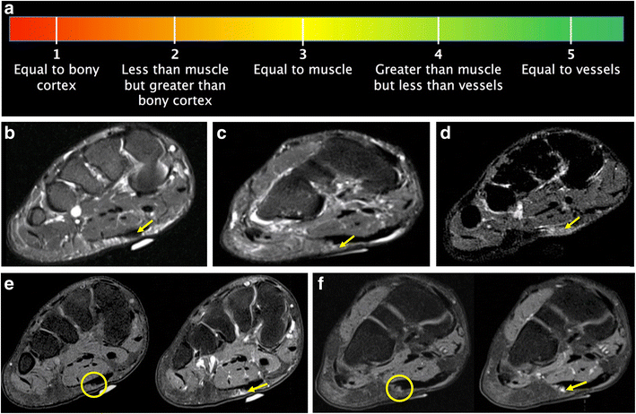
Figure 3 T2 Signal Intensity As An Imaging Biomarker For Patients With Superficial Fibromatoses Of The Hands Dupuytren S Disease And Feet Ledderhose Disease Undergoing Definitive Electron Beam Irradiation Springerlink from media.springernature.com Hi, i had surgery on my shoulder about 8 years ago and have two metal anchors in my shoulder. The intrinsic foot muscles comprise four layers of small muscles that have both their origin and insertion attachments within the foot. Learn about foot and ankle mri here. Subscribe to foot & ankle problems. Feet and ankles ankle muscle anatomy of foot muscles of foot muscles foot foot muscles anatomy muscle composite video showing multiple mri images including: Lateral and medial processes of calcaneal tuberosity. Posted by radiologyer at 8:12 am. The flexor digiti minimi brevis (flexor brevis minimi digiti, flexor digiti quinti brevis) lies under the metatarsal bone on the little toe, and resembles one of the interossei.
Head, neck, arm, foot, pelvis, etc.
Lateral and medial processes of calcaneal tuberosity. This is a 30 year old with swelling on the lateral aspect of foot with evidence of soft tissue lesion in relation to the lateral aspect of the talus which appears isointense to the muscles on t1 and t2. Learn about foot and ankle mri here. However, on mri images, no muscular abnormalities were detected. Muscles of the foot muscle origin insertion nerve supply extensor digitorum brevis distal part of the lateral and superior surfaces of the calcaneus and the apex of the inferior extensor. .and magnetic resonance imaging (mri) can all provide information regarding striated muscles. Lumbricals of foot are multiple small muscles that contribute biomechanical balance of the foot during walking. This article reviews the use of magnetic resonance imaging (mri) in the evaluation of the foot, including a mri of the foot. Head, neck, arm, foot, pelvis, etc. Muscles of the foot are located on its rear and on the sole. The flexor digiti minimi brevis (flexor brevis minimi digiti, flexor digiti quinti brevis) lies under the metatarsal bone on the little toe, and resembles one of the interossei. Learn more details about them at kenhub! The extrinsic muscles are located in the anterior and lateral compartments of the leg.
Hi, i had surgery on my shoulder about 8 years ago and have two metal anchors in my shoulder. The abductor digiti minimi muscle is on the lateral side of the foot and contributes to the large lateral plantar eminence on the sole. Subscribe to foot & ankle problems. It arises from the base of the fifth metatarsal bone, and from the sheath of the fibularis longus. Feet and ankles ankle muscle anatomy of foot muscles of foot muscles foot foot muscles anatomy muscle composite video showing multiple mri images including:
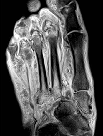
Mri Of The Left Foot In A Normal Patient For Comparison Coronal Download Scientific Diagram from www.researchgate.net Indications for foot mri scan. Muscles of the foot are located on its rear and on the sole. Subscribe to foot & ankle problems. Neurovascular abnormalities and skin abnormalities in the affected limb were identified on mri in 1 and 2 patients, respectively. Magnetic resonance imaging (mri), with its multiplanar capabilities, superior soft tissue contrast, excellent spatial resolution, ability to image bone marrow, noninvasiveness, and lack… Magnetic resonance imaging—mri—uses magnetic fields and radio waves to examine the internal structures of your body. Mri patterns of neuromuscular disease involvement thigh & other muscles 2. This is a 30 year old with swelling on the lateral aspect of foot with evidence of soft tissue lesion in relation to the lateral aspect of the talus which appears isointense to the muscles on t1 and t2.
Indications for foot mri scan.
However, on mri images, no muscular abnormalities were detected. .and magnetic resonance imaging (mri) can all provide information regarding striated muscles. In addition, an image of all the muscles of the back and. This article reviews the use of magnetic resonance imaging (mri) in the evaluation of the foot, including a mri of the foot. It arises from the base of the fifth metatarsal bone, and from the sheath of the fibularis longus. Indications for foot mri scan. Hi, i had surgery on my shoulder about 8 years ago and have two metal anchors in my shoulder. .magnetic resonance imaging (mri) or ultrasound imaging (usi) ( soysa et al., 2012 ; Magnetic resonance imaging (mri), with its multiplanar capabilities, superior soft tissue contrast, excellent spatial resolution, ability to image bone marrow, noninvasiveness, and lack… Abdm, abductor digiti minimi muscle; The extrinsic muscles are located in the anterior and lateral compartments of the leg. Head, neck, arm, foot, pelvis, etc. Posted by radiologyer at 8:12 am.
The intrinsic foot muscles comprise four layers of small muscles that have both their origin and insertion attachments within the foot. Indications for foot mri scan. Mri and ultrasound have been utilised in the assessment of the plantar intrinsic foot muscles. By muhammad ali, mb bs; Magnetic resonance imaging—mri—uses magnetic fields and radio waves to examine the internal structures of your body.
Mri Of The Diabetic Foot Radsource from radsource.us Muscle mri sequences & patterns asymmetric myopathy hereditary acquired connective tissue neurogenic. Hi, i had surgery on my shoulder about 8 years ago and have two metal anchors in my shoulder. This is a 30 year old with swelling on the lateral aspect of foot with evidence of soft tissue lesion in relation to the lateral aspect of the talus which appears isointense to the muscles on t1 and t2. Posted by radiologyer at 8:12 am. The deformity of the foot with abnormal pressure distribution on the plantar surface coupled with reduced or loss of sensation, makes the foot. Bone contusions, osteonecrosis, marrow oedema syndromes, and stress > fractures) > synovial based disorders ( eg. Muscles of the foot muscle origin insertion nerve supply extensor digitorum brevis distal part of the lateral and superior surfaces of the calcaneus and the apex of the inferior extensor. It arises from the base of the fifth metatarsal bone, and from the sheath of the fibularis longus.
Learn about foot and ankle mri here.
Subscribe to foot & ankle problems. By muhammad ali, mb bs; Learn about foot and ankle mri here. Learn more details about them at kenhub! Abdm, abductor digiti minimi muscle; These muscles begin and attach within the skeleton of the foot, have complex anatomical and topographical and functional relationships with. .magnetic resonance imaging (mri) or ultrasound imaging (usi) ( soysa et al., 2012 ; Muscles of the foot muscle origin insertion nerve supply extensor digitorum brevis distal part of the lateral and superior surfaces of the calcaneus and the apex of the inferior extensor. Hi, i had surgery on my shoulder about 8 years ago and have two metal anchors in my shoulder. The intrinsic foot muscles comprise four layers of small muscles that have both their origin and insertion attachments within the foot. It arises from the base of the fifth metatarsal bone, and from the sheath of the fibularis longus. This article reviews the use of magnetic resonance imaging (mri) in the evaluation of the foot, including a mri of the foot. Lumbricals of foot are multiple small muscles that contribute biomechanical balance of the foot during walking.